Speaker
Description
Compared to amorphous silicon ($a$-Si)-based x-ray imaging detectors, the advantage of crystalline Si-based detectors is their fast operation at lower noise, including higher sensitivity owing to larger pixel fill-factor designs. Complementary metal-oxide-semiconductor (CMOS) active-pixel detectors are substituting the conventional $a$-Si for detectors in various imaging modalities. Imaging capability with negligible charge carryover in successive frames derives the CMOS detectors appropriate for dynamic imaging systems in medicine and industry. On the other hand, the CMOS detectors are vulnerable to radiation damage, and the corresponding results are usually manifested as a change in the threshold voltage of transistors and an increase in the dark current of the photodiode within the active pixel, which are responsible for ghosting artifacts and the reduction of dynamic range, respectively [1]. To mitigate the radiation effect, the design of CMOS detectors usually employs an additional optics, called fiber-optic faceplate (FOP), between the x-ray converter and CMOS active-pixel array. The philosophy behind using FOP is that it efficiently transfers light quanta produced by the x-ray converter to the photodiode with minimal loss of light spreading lateral while stopping unattenuated x-ray quanta through the x-ray converter.
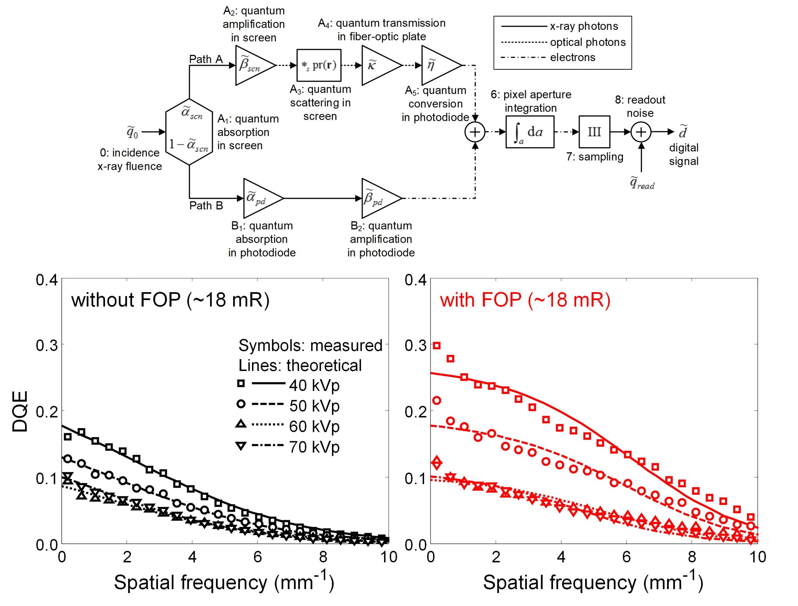
In this study, we investigate the effect of an FOP on the imaging performance of a CMOS detector, such as the modulation-transfer function (MTF), noise-power spectrum (NPS), and detective quantum efficiency (DQE). Stacking a phosphor screen (Carestream Health, Inc., Rochester, NY, USA), FOP (Incom, Inc., Charlton, MA, USA), and CMOS photodiode array (Teledyne Dalsa, Waterloo, ON, Canada) layers completes a prototype CMOS detector. The detector configuration without the FOP layer is the reference. For a range of x-ray energy from 40 to 70 kV, we measure the large-area transfer functions as a function of x-ray exposure (i.e. characteristic curves), MTFs, and NPSs. From these measurements, we calculate the corresponding DQEs.
Briefly, using FOP degrades the MTF performance over entire spatial frequencies, whereas it improves NPS performance and the improvement increases with spatial frequency. We observe that the FOP layer enhances the DQE performance for given energy ranges. To analyze the effect of the FOP layer on the DQE performance, we have developed the cascaded-systems analysis (CSA) model describing the DQE of a CMOS detector employing the FOP layer. The figure attached in this abstract shows the CSA model and compares the measured DQE performances with and without the FOP layers, including the theoretical estimations using the CSA model. We show the measured results with the analysis using the CSA in detail.

[1] D. W. Kim, J. C. Han, S. Yun, and H. K. Kim, “Aging of imaging properties of a CMOS flat-panel detector for dental cone-beam computed tomography,” J. Instrum. 12, p. P01005, 2017.
This work was supported by the National Research Foundation of Korea (NRF) grant funded by the Korea government (MSIP) (No. 2021R1A2C1010161).
Corresponding author: hokyung@pusan.ac.kr