Speaker
Dr
David Hastings
(Christie Hospital)
Description
Background. Molecular imaging using an animal PET camera is a
powerful technique for studying the bio-distribution of
radiolabelled tracers and ligands, offering a means for in-vivo
assessment of new drugs and disease related biochemical processes.
The design of such imaging experiments must be guided by knowledge
of the performance characteristics of the PET camera. For example,
the scientist will need to determine the amount of a compound that
must be administered in order for the radiolabelled molecule to
produce a PET signal with sufficient strength and resolution to
allow detection, visualisation and quantitation. We recently
installed a novel type of animal PET camera, the quad-HIDAC, of
which only three others are installed world-wide (London,
Switzerland, Germany).
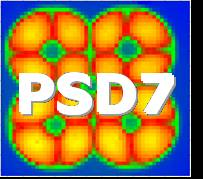
The PET Camera. The HIDAC animal PET system, developed by Oxford
Positron Systems, is based on high-density avalanche chamber
detector modules consisting of argon-flooded multiwire proportional
chambers (MWPC) with integrated converter plates made up of
laminated layers of lead and insulating sheets, drilled with a dense
matrix of small holes, as shown on the left. In each module, the
incoming 511 keV photons interact in a converter plate by
photoelectric and Compton processes to eject electrons into holes
where they are amplified and extracted under a high electric field
gradient into the MWPC. The x-y coordinates of the event are
recorded by the orthogonal cathode tracks. The holes of the
converter planes are 0.4 mm in diameter and 0.5 mm from centre to
centre. Four detector banks, each comprising four HIDAC modules,
surround the imaging port. The axial field of view is 28 cm long and
the transaxial FOV is 17 cm in diameter. Due to the detector design,
the HIDAC system inherently yields information on the depth of
interaction of the photon in the detector banks. Energy information
cannot be obtained with this detector design, but some
discrimination of low-energy photons is inherently performed because
of the reduced sensitivity of the detectors to low-energy gammas.
Results. The following performance parameters were determined. The
values of mean spatial resolution in the three spatial planes were
1.02mm in the horizontal direction, 1.00 mm in the vertical
direction, and 0.97mm in the axial direction. Furthermore,
measurements across the field of view showed these resolution
results to be invariant within a standard deviation of 0.05mm.
Therefore, the volumetric resolution is 1.0 cubic mm, or 1
microlitre, which is better than for any other PET system. Absolute
sensitivity (scatter-corrected) measured with a point source was
found to be 0.75%. Count-rate capability measurements showed a near-
linear response at low activities, rising to a 20% loss rate at an
activity of 11.5MBq. These results will be essential in designing
animal imaging studies for in vivo assessments of new compounds and
biochemical processes. A preliminary study using [18-F]-
Fluorodeoxyglucose (FDG), shown on the right, clearly demonstrates
superb detail of the in vivo tissue biodistribution of glucose
metabolism, with muscles, heart, and individual vertebrae
musculature being well defined.
Conclusion. This PET camera was found to have better spatial
resolution and better uniformity of response over a larger field of
view than has previously been reported, and should allow excellent
imaging assessment of small regions of radiotracer uptake in the
mouse.
Primary author
Dr
David Hastings
(Christie Hospital)
