MEDAMI 2016 - IV Mediterranean Thematic Workshop in Advanced Molecular Imaging

Europe/Zurich
Description

Vision Paper on Medical Imaging

and ZIP file with photos of the workshop

at the bottom of this page

Personalized Medicine, a Paradigm Shift for Medical Imaging?

Towards Personalized Medical Imaging: How to implement the vision

Ajaccio Bay, Corsica, May 1-5, 2016

Porticcio bay 


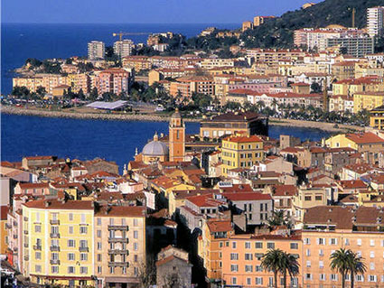
Following the series of previously conducted symposia on dedicated medical imaging instrumentation, we are presently organizing the 4th issue of this workshop to be held on the beautiful island of Corsica.

The purpose of this meeting is to discuss the implications of the vision of individualized/personalized medicine on the activity and productivity of the imaging instrumentation community. This activity needs to be in tune with the future demands of the personalized  medicine.

The key motivation for the personalized medicine is to deliver the correct treatment to the particular patient at the right time, while controlling the overall costs of providing healthcare to the public. A key requirement of our future healthcare system is the capability to provide more effective preventive screening care to the ageing population to reduce the incidence of patients presenting with late-stage disease and the associated high costs in managing the medical care for these patients.

Some of the highlight topics of this meeting are:

  • Personalized Medicine – what will be the impact on current medical imaging?
  • Is there a need for organ specific (precision) medical imaging?
  • What are the medical areas with the strongest impact/needs?
  • New Imaging Technologies and Methods for precision imaging, including image guided radio- and HIFU-therapy and drug delivery.
  • The role of new tracers and agents in personalised medical imaging.
  •  Funding mechanisms for the development of dedicated imagers, from development to implementation, the role of government agencies and private investment.

Preliminary list of key speakers at this meeting include:

  • Ruxandra Draghia-Alkli, Head of the Health Directorate at the EU DG Research
  • Prof. Roderic Pettigrew, director of the National Institute of Biomedical Imaging and Bioengineering (NIBIB)
  • Magda Chlebus, Head of strategy at the EFPIA (European Federation of Pharmaceutical Industries and Associations)
  • Prof. David Townsend, National University of Singapore, inventor of PET/CT
  • Prof. Osman Ratib, Head of the Nuclear Medicine department, University Hospital Geneva
  • Prof. John Prior, Head of the Nuclear Medicine department, University Hospital Lausanne
  • Prof. Markus Schwaiger, Head of the Nuclear Medicine department, Klinikum Rechts der Isar, Münich
  • Prof. Nassir Navab, Computer Aided Medicine, TU Münich
  • Prof. Craig Levin, Stanford Molecular Imaging Instrumentation Laboratory (MIIL)

The meeting format fosters a close interaction between different stakeholders from academia, medical institutions and organizations, regulatory agencies and industry. In particular, industry is invited to play an active role by:

  • Giving keynote lectures on the vision of industry towards personalised medicine.
  • Actively participating in sessions and round table discussions.
  • Meeting with government agencies’ representatives to express industry position.
  • Conducting one-on-one meetings with key researchers in the field.

The list of partners having a role to play in this broad discussion includes, but is not limited to: scientific community, medical community, high-tech industry, medical companies, pharmacological companies, government agencies, local governments, foundations, patient support organizations, health insurance organizations and companies, media, etc. Therefore, we would like to invite representatives of these groups to the “discussion table”. Based on our historically documented natural scientific interest in medical imaging, we perceive ourselves as potentially natural instigators of such a discussion.

Hence, we are inviting representatives of the stakeholders and other participants in this process to a four-day meeting in Ajaccio, Corsica, May 1-5, 2016. The venue and the format of the meeting (that are both being finalized at this moment) will be to facilitate the multi-partner multidisciplinary discussions in a non-disruptive enabling atmosphere with several round table brainstorming sessions, in addition to the invited talks by experts and representatives in the relevant fields and subjects, focusing on different selected aspects of the overall process including science, medicine, industry, and with points of view of decision makers’ developing healthcare policies also included in this discussion. 
 

P. Lecoq, CERN, Geneva, Switzerland

J.M. Benlloch, I3M, Valencia, Spain

F. Garibaldi, ISS&INFN, Rome, Italy

Y. Hämisch, Axel Schröder Unternehmensberatung GmbH & Co. KG

C. Levin, Stanford, USA

G. Loudos, Technological Educational Institute, Athens, Greece

S. Majewski, University of Virginia, USA

D. Townsend, A*STAR-NUS Clinical Imaging Research Centre, Singapore

V. Sossi, University of British Columbia, Vancouver, Canada


---------------------------------------------------------------------------------------------------------------------------------------

Our Platinum Sponsors

Philips CERN logo


Our Gold Sponsors


Our Silver Sponsors


Our Bronze Sponsors

---------------------------------------------------------------------------------------------------------------------------------------

Supporting Organisations

CERN, COST ACTION TD1401 FAST, CRYSTAL CLEAR, EANM, EIBIR, ERC TICAL #338953 , ESMI, IEEE, MINDVIEW, TRIAGE


Participants
  • alberto Del Guerra
  • Alberto Di Meglio
  • Alessio Ghezzi
  • Alexandre Lombardo
  • Alexis Moscoso Rial
  • Antonio J. Gonzalez Martinez
  • Benedikt Wiestler
  • Bengt Langstrom
  • Bernd Pichler
  • Carlos Correcher
  • Catherine Cheze Le Rest
  • Charalampos Tsoumpas
  • Christian Morel
  • Craig Levin
  • Daniel Gagnon
  • David García-Juan
  • David Sanchez
  • Dennis Schaart
  • Dimitris VISVIKIS
  • Dominique Thers
  • Dominique Yvon
  • Elena Popova
  • Eric Mas
  • Etiennette Auffray
  • Felicie Sherer
  • Ferid Haddad
  • Francesco Scopinaro
  • Franco Garibaldi
  • Gabriela Llosá
  • Giacomo Traini
  • Gérard Tauzin
  • Homer Pien
  • Jesús Silva Rodríguez
  • John Prior
  • Jose Benlloch
  • Luc Bidaut
  • Magda Chlebus
  • Manuela Cirilli
  • Marco Paganoni
  • Marco Pizzichemi
  • Markus Ehrenfried
  • Markus Schwaiger
  • Maurizio Conti
  • Michael Seimetz
  • Nassir Navab
  • Olga Kochebina
  • Osman Ratib
  • Pablo Aguiar
  • Paul Lecoq
  • Piergiorgio Cerello
  • Radek Zboril
  • Ricardo Graciani Diaz
  • Roderic Pettigrew
  • Rosana Martinez Turtos
  • Rouslan Sitnikov
  • Ruxandra Draghia Akli
  • Samo Korpar
  • Sebastian Aussenhofer
  • Sergei Dolinsky
  • Sergey Vinogradov
  • Stefaan Tavernier
  • Stefaan Vandenberghe
  • Stefan Gundacker
  • Stephen Bartold
  • Veronica Regazzoni
  • Vincent Czajkowski
  • William Moses
  • York Haemisch
    • Precision Medicine – vision, concept, stakeholders and plans for implementation: Chair: P. Lecoq, CERN, CH

      Chair: P. Lecoq, CERN

    • 15:45
      Coffee break
    • Precision Medicine – vision, concept, stakeholders and plans for implementation: Chair: P. Lecoq, CERN, CH

      Chair: P. Lecoq, CERN

    • Imaging for minimizing interventions: Chair : L. Bidaut, Northeast Scotland, UK
      • 6
        Nuclear Medicine and Molecular Imaging for Minimizing Medical and Surgical Interventions
        This presentation will show actual and upcoming applications of nuclear medicine and molecular imaging for minimizing medical and surgical interventions. There are several ways nuclear medicine and molecular imaging can do this: (1) early detection of potentially lesion (rule-in/rule-out); (2) easier targeting of biopsies; (3) better detection of micro-metastases and tumor margins during oncological surgery; (4) help in deciding minimal invasive, standard surgical, or watch-and wait approaches; and (5) early detection of residual disease or recurrence, among others.
        Speaker: J. Prior, CHUV Lausanne (CHUV University Hospital)
      • 7
        Precision Surgery with a novel radio-guided surgery
        Speaker: G. Traini, Sapienza Univ. Roma
      • 8
        Robotics and Augmented Reality for Patient and Process Specific Imaging and Visualization
        Speaker: N. Navab, TU Münich
    • Theranostics: Chair : M. Schwaiger, TU Münich, Nuclear Med., De
      • 9
        Theranostics: a key component of personalised medicine
        Speaker: O. Ratib, HUGE, Geneva
      • 10
        Prostate cancer (PCa) : A Theragnostic evaluation of radioisotope methods
        Prostate cancer is the most common malignancy among males of developed countries. Several methods of diagnosis, staging and therapy have been recently studied : among them radio isotope methods are growing and probably are helping personalization and tailoring of treatment. Tailoring is important in PCa because High and low risk, neuroebdocrine or glandular, hormone therapy responsive or castration resistant ( CR) PCa need different cure and also different attention and management. New specific pharmaceuticals as well as therapeutic radio-pharmaceuticals, e.g. the bone seeking alpha emitters such as the relatively new 223RaCl2 , or also some beta emitter agents, can prolong the survival of patients by curing a selected type or site of lesion, e.g. only bone metastases from castration resistant PCa. We need accurate methods of Node (N) / Distant metastases (M) staging and of biological characterization to select the patients and plan the exact therapy. Non invasive imaging methods include contrast enhanced CT and NMR, diffusion NMR and echography. These imaging methods are of great help in the management of patients with PCa, but echography has a very limited field of view and despite substantial technological progress , CT and NMR are not enough sensitive nor specific to become reliable standard methods to detect nodes and to guide surgical or radiotherapic cure . 18F/11C Choline (FCH) PET is specific but not highly sensitive for Tumor( T) and N staging, the accuracy of FCH for early bone metastases is controversial. Experiences conducted by our group showed high accuracy of 99mTc Bombesin SPECT for N staging. Our data have recently been confirmed by Mistakis et al, who detected higher uptake of 68Ga MJ9 - a bombesin analogue- than FCH in PCa invaded nodes. Bombesin is a growth factor for androgen dependent and also androgen independent PCa, because it can be secreted also by PCa cells that underwent neuroendocrine shift. Our group was able to demonstrate this circumstance in some- though few- patients by using neuroendocrine seeking and glandular PCa seeking radiopharmaceuticals, such as 111In somatostatin and99mTc bombesin or 18F FCH and 68Ga DOTANOC. In these patients the PCa specific therapy can be integrated. HPED-CC, a PSMA inhibitor, will also play an important role in a near future as a PCa seeking agent and can be used for diagnostic use when labeled with 68Ga and for therapy when labeled with 177Lu. Early diagnostic trials with 68Ga HPED-CC have recently shown accuracy as high as 95% in detecting relapsed nodes. Theragnostics means coordinated procedures closely linking diagnostic and therapeutic methods in order to selectively cure the patients, in other words to personalize the patients' management. As a conclusion we can observe that new radiopharmaceuticals, though at the moment still used for clinical research or very recently become costumer available can give a substantial contribution to personalize/tailor the therapy of PCa patients as theragnostic novel procedures . Aknowledgements: We are indebted with John Prior and the team of Lausanne CHUF for their help on 68Ga MJ9
        Speaker: F. Scopinaro, Sapienza Univ. Roma1 (Sapienza University of Rome)
      • 11
        Radioisotope production and the Arronax Facility
        Radioisotopes can be used in different field of medicine like oncology, neurology and cardiology. Different types of applications are possible thanks to the different kind of radiation available through the radioactive decay of nucleus. Gammas which are penetrating radiation are used for imaging to help diagnosis whereas charged radiations are used for therapy to destroy cells. Only few radionuclides can be used directly (I131 for thyroid cancer is one example). In most cases, these radionuclides must be coupled to a carrier molecule (a vector) to target the cells of interest. This labelled vector forms a radiopharmaceutical. A vector can be a chemical molecule, a peptide or an antibody and its distribution time in the body is dependent on its size. Peptide can distribute within hours whereas antibodies need days. Currently, only few isotopes are used in clinical practice (Tc99m, F18 for imaging and I131 and Y90 for therapy). However, many others may be of medical interest due to their emitted radiations (alpha emitters, Auger emitters) and / or their half-lives that can be adapted to the carrier molecule transit time and to the pathology. Recently, with the recent technological advances, it is possible to combine imaging information and therapeutic use of radionuclides which is called the theranostic approach. This approach allows personalizing the treatment to each patient. The diagnosis test done prior to the treatment allows following and controlling the patient response to the injected radiopharmaceutical. It allows a better control of the targeting and increases the benefit/toxicity ratio as useless treatments on patients with no response to the diagnosis test are avoided. For the theranostic approach, it is preferable to use pairs of radioisotopes of the same element like (I124/131I, Cu64/Cu67, ). All these points lead to a renewal interest on isotope production and the necessity to have dedicated facilities like the Arronax facility in France which is devoted to the production of innovative radionuclides for medical applications.
        Speaker: F. Haddad, Subatech & ARRONAX (Subatech and GIP ARRONAx)
      • 12
        Personalised production of medical radioisotopes with laser-accelerated protons
        Many diagnostic methods are based on the use of tracers labelled with radioactive isotopes. Their production in centralised facilities and delivery to local health centres imply strong constraints to the isotope half lives. For this reason, more than 90% of all PET interventions are based on F-18 at present. The on-site synthesis of short-lived tracers containing C-11, O-15, or N-13 would allow for a wealth of drugs with ideal characteristics for each patient and pathology. The production of radioisotopes requires beams of accelerated protons or deuterons with energies around 15-20 MeV/u. A novel acceleration technique based on highly intense, pulsed lasers has the potential to provide such beams at much lower cost than classical synchrotrons. We present the development of a dedicated setup aiming at the production of PET isotopes, comprising a table-top, terawatt laser with high repetition rate. With this setup we have recently achieved the first demonstration of laser-proton acceleration in Spain. In addition, we have calculated the requirements for the synthesis of useful quantities of different isotopes in various reaction channels.
        Speaker: M. Seimetz, I3M Valencia (Instituto de Instrumentación para Imagen Molecular (I3M))
    • 10:00
      Coffee Break
    • Theranostics: Chair : M. Schwaiger, TU Münich, Nuclear Med., De
      • 13
        Prostate-Checker: Prostate cancer assessment by multi-parametric MRI studies
        Prostate cancer (PC) is the second most diagnosed type of cancer and the fifth leading cause of cancer-related death in men worldwide (most frequent cause of cancer death in men in developed countries). Guidelines about prostate magnetic resonance (MR) imaging published by the European Society of Urogenital Radiology (ESUR) in 2012 recommended a multi-parametric approach for a better characterization of PC. Available sequences that allow the acquisition of anatomical and functional studies have led MRI to be the modality of choice in PC evaluation and during follow-up studies. Anatomic T2-weighted (T2W) images, diffusion-weighted (DW) images and dynamic contrast enhanced (DCE) series allow the assessment of interstitial edema, cellularity and micro-vascularity of the gland respectively. MR imaging derived biomarkers provide quantitative information to objectively characterize a pathological process or a therapeutic action. A software prototype (Prostate Checker Ltd, UK http://prostatechecker.co.uk) is presented (Figure 1). The tool is capable of performing voxelwise multi-parametric analysis from T2W, DW and DCE MR images to extract several imaging biomarkers related to PC detection and grading. Imaging biomarkers and their multi-variate combination are displayed in the form of parametric maps (Figure 2). As images have different spatial resolutions and space orientation, and the prostate may slightly change in position, the software performs a re-slicing and elastic co-registration, driving all images to a common reference space and resolution. Once the spatial coherence is achieved, the user manually segments the prostate or any PI-RADS region, launching the complementary multi-parametric analyses based on T2W, DW and DCE images. First module of the prototype applies advanced TexRAD texture analysis (licenced by TexRAD Ltd www.texrad.com, part of Feedback Plc) to T2W images to quantify tissue heterogeneity through a filtration-histogram technique. First step uses a band-pass Laplacian of Gaussian (Mexican hat shaped filter similar to a non-orthogonal Wavelet approach) to extracts and enhances texture features of different sizes corresponding to spatial scale filter (SSF). Second step performs histogram-analysis to describe the shape of the histogram e.g. mean intensity/mean of positive pixels, standard-deviation, entropy, kurtosis and skewness. Diverse published literature states the use of filtration-histogram texture analysis technique to assist in risk-stratification. A second module applies pharmacokinetic models to the DCE series to characterize tissue micro-capillarity. This module extracts and displays parameters such as the transfer constant (Ktrans), the reverse transfer constant (кеp) and the extracellular space fractional volume (ⱱе), widely reported in literature to have a high sensitivity in cancer detection. The third module exploits DW images computing and displaying apparent diffusion (ADC) maps and intra-voxel incoherent motion (IVIM) parameters when several b-values are acquired. PC shows a lower ADC and D properties, with higher D* and f properties. Nosologic images from the combination of the different extracted biomarkers are created by their combination through the application of multivariate analysis, providing closer information to the clinical endpoints.
        Speaker: D. García-Juan, Quibim SL (Quibim SL)
      • 14
        Imaging the continuous spectrum of therapeutic radionuclides with detectors and pinholes suited for high energy
        Targeted radionuclide therapy (TRT) is an established cancer treatment modality. It relies on cancer specific agents that are labeled with radionuclides for internal radiotherapy. The biological effect to tissues is generated by the energy absorbed from the radiation (typically beta-emittors) emitted by the radionuclide, TRT can result in substantial sparing of uninvolved tissue and organs and therefore avoid adverse events compared to conventional external beam therapy. Current PET or SPECT systems are suboptimal for imaging isotopes used in radionuclide therapy. SPECT (developed for single gamma photons with mono-energetic low-energy emission) has been used as imaging that has very limited use in the specific domain of radionuclide therapy due to poor quantification. PET can only be used for Y-90, which has a small fraction of positron emission. The main limitations of current SPECT systems for these isotopes are related to the high penetration in the collimator and low detection efficiency of the used detectors. Secondary bremsstrahlung photons, characterised by a continuous energy spectrum, up to the maximum energy of the emitted electron, need a collimator with minimal penetration similar to high-energy pinhole SPECT. To efficiently detect incoming photons, dense high-energy detectors similar to those used in PET should be used. The conventional 3/8 inch NaI works well at low energies but has only very limited stopping power at 1 MeV. The currrent standard PET scintillator L(Y)SO is not suited for this task due to its high intrinsic activity (which cannot be removed when acquiring in singles mode). BGO is more suited, the main advantage of BGO comes from the high amount of direct photo-electric interactions and resulting smaller amount of Compton interactions arriving in the lower energy window. A disadvantage of BGO is the smaller amount of scintillation light resulting in reduced energy resolution at lower energies (>30% at 100 keV and 15 % at 1 Mev), but given the continuous spectrum this is less important. As the spectrum from a typical therapeutic radionuclide will be mostly composed of low energies, a relatively thin detector can be considered (about 1 cm was selected from simulation study). Based on the expected resolution of these detectors a stationary system can be designed with 1 cm spatial resolution in the cFOV.
        Speaker: S. Vandenberghe, Ugent (Ugent)
      • 15
        Nanotechnologies in diagnostic and theranostic applications

        Nanomaterials based mainly on polymer nanostructures, magnetic nanoparticles and carbon nanoallotropes represent challenging solution in various diagnostic, therapeutic and theranostic applications [1-6]. The present contribution explores the use of superparamagnetic nanoparticles as contrast agents in MRI diagnostics and theranostics involving the results of clinical trials. Various types of polymer and magnetic carriers used in targeted drug delivery are compared in terms of the drug loading and drug release mechanisms. The possibilities of carbon nanostructures (nanodiamonds, carbon nanotubes, graphene derivatives, carbon dots) and their hybrids in photoluminescent imaging, combined magneto-fluorescent imaging and drug delivery are also summarized. The specific attention is focused on photoluminescent carbon dots, control of their optical properties, toxicity and biodistribution. Their use for selective cell labeling, photoacoustic imaging, photodynamic therapy and targeted drug delivery is analyzed taking into account their emission characteristics, surface chemistry and structural properties.

        Speaker: Prof. R. Zboril, Olomuk Univ. (Cz) (Regional Centre of Advanced Technologies and Materials, Department of Physical Chemistry, Faculty of Science, PalackýUniversity Olomouc, Tř. 17. listopadu 12, 771 46 Olomouc, Czech Republic)
    • Data Mining: Chair : P. Lecoq, CERN
      • 16
        Machine Learning: recent developments and future impact on medical science and technology
        Speaker: N. Navab, TU Münich
      • 17
        Big data management - From CERN/LHC to personalised medicine
        The transformations that have taken place in Information and Communication Technology in the past 20 years have given rise to a new form of scientific research paradigm where data-intensive, large-scale projects combine experiment, theory and computing to address fundamental questions about ourselves and our universe. The large-scale computing and data analysis infrastructure set up by the High Energy Physics community to support the research of the LHC Experiments at CERN and in the hundreds of collaborating facilities worldwide is one of the foremost examples of this paradigm. Today the HEP community is not anymore the only place where increasingly large amounts of data are produced. Biomedical and healthcare research and practice could benefit from a broader use of big data analysis and simulation platforms. However, the biomedical domain requires a new focus on careful governance and use of data and information in the respect of the social and human value of such data and the design and deployment of collaborative frameworks where medical research, clinical practice and modern information technologies can constructively interact with each other to deliver personalized care. This talk briefly describes the state of the art of large-scale data analytics platforms as used in the HEP community and the ongoing work to adapt and extend such platforms for the benefit of medical applications.
        Speaker: Alberto Di Meglio (CERN)
      • 18
        Multi-parametric multi-modality imaging for prognostic and predictive modeling in oncology
        Speaker: D. Visvikis, INSERM France
    • 12:30
      Lunch break
    • Industry: Chair : Y. Haemisch, LDTEC Consulting, De
      • 19
        On the role of imaging and data analytics in personalized medicine and population health: a view from Philips
        Healthcare around the world is undergoing dramatic transformations, due primarily to the unsustainable rate of growth in the cost of care. In response, the community must provide better care at a lower cost. Personalized medicine plays an integral part in that. At Philips we are pursuing a strategy in precision health, which links across the entire care continuum from monitoring of healthy living, to early and definitive diagnosis and prevention, to more precisely therapies, to enabling safer transitions from hospital to home-based care, and ultimately to healthy living again. Imaging plays a central role in rendering definitive diagnosis and therapy selection. As a biomarker, imaging is needed for localization and staging of many diseases; it can be used to effectively segregate cohorts of patients by prognosis, and it can help select therapies, guide interventions, and monitor the effectiveness of therapies. Significantly, imaging – with its tissue-level view of the patient – wields even greater diagnostic power when combined with cellular-level views of the patient through histopathology, as well as the molecular level view through genomics, molecular pathology, and other ‘omics. To succeed, we need to apply analytics techniques to both extract greater information from each modality, as well as to establish causality between modalities operating at different scales. The potential to extract greater information content is perhaps more pronounced with PET than any other imaging modality. Introduction of the digital PET (dPET) provides us with unprecedented resolution and CNR, which will likely translate into earlier diagnosis and greater quantification of disease. Yet evidence for improved diagnostic and prognostic value derived from these improvements are still accrued at present, especially in the context of oncology, neurology, and cardiac patients. The use of PET during interventional therapies remains largely unexplored at this time. PET can likely also play a greater role in producing dose maps of chemoembolization therapies. Additionally, the ability of PET to offer physiological and metabolic insights will likely be an important complement to cellular and molecular characterizations of disease. In short, we believe that imaging in general, and molecular imaging in particular, needs to heed the call for value-based transformation of healthcare. The implication is that, we, as a community, needs to focus on providing greater clinical value in our imaging studies, and developing techniques which are easier to use, protocols which are more repeatable, and findings which are more quantitative and more indicative of pathological or physiological changes in patients.
        Speaker: Homer H. Pien, Phillips (Philips)
      • 21
        Form High-energy physics to medical applications.
        CERN is one of the world's best centres for fundamental research. However, the economic return has been disappointingly low. I have been active in the field of technology transfer from fundamental research in high-energy physics to other fields during the last 20 years, and this activity culminated in the creation of a spin-off company a few years sago. I will analyse the reason for the low economic return of such fundamental research, on the basis of my personal experience over the last 20 years.
        Speaker: S. Tavernier, PETSys (Vrije Universiteit Brussel (BE))
      • 22
        Technology Transfer at CERN
        Speaker: M. Cirilli, CERN
    • 15:40
      Coffee break
    • Brain (including open MINDVIEW session): Chair : J.M. Benlloch, I3M, Sp
      • 23
        Molecularly targeted therapy and radiogenomic imaging in glioblastoma
        Glioblastoma (WHO grade IV) is the most common malignant primary brain tumor in adults with a dismal median overall survival of 16 months, despite intensive radio-chemotherapy. In recent years, the advent of high-throughput genomic analyses has helped us to better understand the biology underlying this disease. These advances translate in two ways “from bench to bedside”: First, tumors now can robustly be grouped in molecularly defined subgroups, which increasingly complement the WHO classification, and even prove to be prognostically superior to it. Secondly, key molecular “driver” alterations and biomarkers predictive of therapy response have been identified and caused a great interest in the development of molecularly targeted therapies in these highly treatment-resistant tumors. Today, treatment decisions are increasingly based on defined molecular biomarkers, most prominently the methylation status of O6-methylguanine-DNA methyltransferase (MGMT) promoter or combined deletion of the short arm of chromosome 1 and the long arm of chromosome 19 (1p/19q codeletion), a hallmark of oligodendroglial tumors. Furthermore, several actionable alterations have been identified which are recurrently found in these tumors, such as isocitrate dehydrogenase (IDH) mutations, epidermal growth factor reception (EGFR) amplifications or FGFR-TACC fusions. In parallel, fostered by advancements in MR and PET imaging and post-processing, the field of radiogenomics, investigating how genomics are reflected in the imaging phenotype, has received increasing attention. These developments have been met with great enthusiasm, as drugs targeting defined genomic alterations promised to improve therapeutic options for this disastrous disease, while hopefully reducing side effects compared to conventional chemotherapeutic agents.
        Speaker: B. Wiestler, Dept. of Neuroradiology, TU Munich (Dept. of Neuroradiology, TU Munich, Germany)
      • 24
        The TRIMAGE project: A Trimodality brain scanner for early diagnosis of schizophrenia
        TRIMAGE is an interdisciplinary FP7-funded European collaboration aimed at developing a cost-effective dedicated brain PET/MR/EEG brain scanner for early diagnosis of schizophrenia. The brain activity measured with fMRI, combined with the highly sensitive molecular information provided by PET, and the highly sensitive temporal information from EEG converge into a new imaging tool for diagnosing, monitoring and follow-up of mental disorders. As for the clinical aspects we are interested in the multimodal assessment of response inhibition. The Loudness Dependence of Auditory Evoked Potential (LDAEP) is a suitable biomarker of inhibitory action in signal processing. Patients with schizophrenia may exhibit alterations in the responsiveness to sensory stimuli (i.e., stronger LDAEP values). We aim to further elucidate the relationship between multimodal neuroimaging methods and dimensions of symptoms, observable behavior, personality traits and general psychopathological dysfunction. A sample of 20 healthy controls and 20 patients with manifest schizophrenia will be initially examined with the LDAEP paradigm in a trimodal approach with the available 3T MR-PET scanners in Munich and Jülich. In Munich, FDOPA will be used and static and dynamic analyses will be compared with fMRI data; in Jülich, PET measurements with the radiotracer [11C]-flumazenil will assess the binding potentials of GABA-A receptors. MRS will provide data about GABA concentrations. At the end of this first clinical evaluation a set of suitable biomarkers will be proposed. The MR magnet will be cryogen-free with main B0 field of 1.5 T. Magnet warm bore is 720 mm with a field uniformity of ±1 ppm and a field stability <0.1 ppm/hour. The 5 gauss line fringe field will be < 2.8 m axially. The gradient coil has a maximum strength of 42 mT/m (X,Y) and 41.2 mT/m (Z) with a slew rate of 123 T/m/s (X,Y) and 127 T/m/s (Z). The typical MR sequences to be uses will be: UTE (for attenuation correction), MPRAGE and FLAIR (for anatomical information) EPIK (for High resolution functional information) The PET component is designed to provide performance beyond the state of the art for clinical PET systems with an expected spatial resolution of about 2 mm FWHM. The PET field-of-view will be 162 mm axially and 240 mm diameter with an open bore of 308 mm diameter. The PET detector comprises 216 tiles featuring two layers of LYSO crystal matrices (3.4 mm pitch) with half pitch staggering. SiPM matrices will be used as photodetectors, and a DAQ based on the TRIROC ASIC and FPGAs will take data in list mode. A state-of-the-art MR-compatible EEG cap with 32 channels will be simultaneously used with the PET/MR scan. The results of the first clinical results, of the simulations and of the experimental tests will be presented “The research leading to these results has received funding from the European Union Seventh Framework Programme (FP7/2007-2013) under grant agreement n° 602621- Trimage.“
        Speaker: A. Del Guerra, Dept. of Physics, University of Pisa (Dept. of Physics, University of Pisa)
      • 25
        SPM-guided PET analysis for the evaluation of non-lesional epilepsy.
        Multi-modal preoperative evaluation with advanced structural, functional, and metabolic neuroimaging techniques is essential in the pre-surgical evaluation of refractory epilepsy for the delineation of the epileptogenic zone to be resected. In latter years, PET has gained a leading role in this evaluation since it has demonstrated to be simpler and more sensitive than ictal SPECT in certain situations. Furthermore, the availability of co-registered PET/MRI images has improved the interpretation of images of both modalities and enabled a more straightforward use of PET information on MRI-guided surgery. In this regard, MRI findings have demonstrated to be extremely useful for assisting PET analysis, since the location of anatomic anomalies related with epilepsy can focus the PET analysis on a reduced area, making possible to detect small focal hypometabolisms that might be dismissed on a simple visual inspection. Nevertheless, 35% of epilepsy patients show no lesions on the MRI, which are related with an earlier stage of the disease, and resective epilepsy surgery in non-lesional epilepsy is commonly associated with less favourable outcome. For these patients, PET is of particular importance, but its performance is reduced due to the lack of the aforementioned MRI guiding. In this context, the use of PET quantification techniques, such as Atlas-based Asymmetry Indices (AI) and especially Statistical Parametric Mapping (SPM) has improved the localization of the epileptogenic focus. In particular, SPM has potential for being a suitable substitute of MRI guiding the PET analysis when no anatomical lesions are found. On this work we evaluated the correlation between anatomical findings and SPM results on 24 patients with different types of epilepsy-related MRI lesions (12 cortical focal dysplasia, 12 mesial temporal sclerosis) and 5 patients with non-lesional MRI. PET images were processed with iSFS-RR resolution recovery algorithms and SPM maps were obtained by an unpaired t-test voxel-by-boxel comparison between the patient and the database of 97 healthy patients. Patients were evaluated by using PET, PET/MR and PET/SPM images, trying to reproduce a clinical scenario. On lesional epilepsy patients, SPM provided better sensitivity (91.6%) than PET only images (70.8%), and SPM findings showed high correlation with MRI anatomical findings. When applied to non-lesional epilepsy patients, PET/SPM also offered better sensitivity (80%) than simple PET visual analysis (40%). Thus, the purposed SPM-guided PET visual analysis demonstrated to be more effective than the routine visual inspection, showing potential for improving focus location on non-lesional epilepsy.
        Speaker: J. Silva-Rodríguez, IDIS Santiago de Compostela (Molecular Imaging Group in Health Research Institute (IDIS), Santiago de Compostela, Galicia, Spain)
      • 26
        Final design and initial results of the first MINDView brain PET insert prototype
        The first prototype of the MINDView project, a brain PET insert MR compatible, is currently being assembled. The scanner is composed of 3 rings of 20 detector blocks each. The detector block includes a monolithic LYSO crystal with 50x50x20 mm3 and a custom 12x12 SiPM array (TSV-type). The system defines an axial and transaxial field of view (FOV) of about 150 mm and 240 mm, respectively. Detector blocks are kept at a stable temperature in the range of 20-25ºC using controlled temperature air cooling. The X and Y light projections of each detector are measured and from them the planar and depth of interaction (DOI) positions deduced. Here, several methods have been studied namely traditional Center of Gravity (CoG), Rise to Power (RTP), Fitting profiles to the light distribution but also Neuronal Networks. Using the standard approaches (CoG and RTP) it has been possible to characterize the DOI with a resolution of about 5 mm. This makes it possible to reach an average detector spatial resolution (without source finite size corrections) of about 2.6 mm for the whole crystal volume, improving to 1.7 mm, at DOI’s values closer to the photosensor. Average energy resolution ranging from 17% at the crystal entrance down to 16% near the photosensor, is obtained. Parts of the detector ring have been successfully tested for RF shielding and eddy currents. This was carried out by using a Radio frequency (RF) screen structure based on carbon fiber composites with specific thickness and orientations.
        Speaker: A. Gonzalez Martinez, I3M Valencia
      • 27
        MINDVIEW open session, ctn'd
        Speaker: J.M. Benlloch, I3M Valencia
    • Overview Molecular Imaging Technologies: Chair : W.W. Moses, LBNL, USA
      • 28
        Past, Present and Future of Positron Emission Tomography
        Positron Emission Tomography (PET) is a well established imaging technique for in vivo molecular imaging. After a brief history of PET, the physical principles and the main performance parameters are presented. The evolution of the technology that has brought PET from a bench experiment to a clinical indispensable instrument is fully illustrated. In particular, the present limitations and the expected future performance of the PET tomographs are discussed, both as for the hardware and software aspects. The status of art of clinical, preclinical and hybrid scanners (i.e., PET/CT and PET/MR) is shown. Finally the recent and future technological developments are presented. As a specific example, the current applications of PET to range monitoring in particle therapy are discussed.
        Speaker: A. Del Guerra, Dept. of Physics, University of Pisa, Italy (Dept. of Physics, University of Pisa, Italy)
      • 29
        Nuclear and CT imaging: what drives it ?
        Since the invention of the CAT scan by Hounsfield in the early 1970s, transmission and emission imaging modalities used in radiology and nuclear medicine have continually benefit from improvements in detection technology, signal treatment and applied mathematics. The development of 3D PET in the late 1980s, and of positron rotating partial-ring tomographs leaving potentially enough void between arrays of detectors to insert an X-ray tube and X-ray detectors, led to the invention of PET/CT in the early 1990s. Although the original concept of PET/CT could not be implemented as envisaged, the advent of the first PET/CT prototype in the late 1990s, which provided native fusion of anatomical and functional images, revolutionized rapidly both the practice and the market of nuclear medical imaging. Riding the wave of PET/CT that was claimed to be the invention of the year 2000 by the Time Magazine, and thanks the development of solid-state photodetector devices insensitive to magnetic fields, industry majors have made recently a determined effort to bring PET/MR devices to the market. Although the pertinence of this new evolution of the imaging discipline remains to be assessed, it is nevertheless obvious that every technological breakthrough that would bring better insight and quantification of the metabolic function, or of several molecular pathways at a time, together with precise anatomical information, possibly at a reduced absorbed dose, constitutes a genetic trend for the development of future hybrid nuclear and CT imaging modalities. In this regard, the invention of massively parallel hybrid pixel detectors for charged particle trajectography in high energy physics, once applied to the detection of X-rays for photon counting CT, paves the way to the development of spectral CT that will potentially provide the first ever intrinsically anatomo-functional medical imaging device, which could be used to image several functionalized contrast agents made of nanoparticles. In a similar line, improvements of scintillation spectrometry implementing photonic devices and novel compact and fast photodetectors such as SiPMs permits to improve dramatically signal-to-noise ratio of medical images by exploiting TOF information in the image reconstruction process. Hence, extrapolating this trend may possibly foreshadow the advent of reconstructionless positron tomography in some unknown but brilliant future of nuclear and CT imaging.
        Speaker: C. Morel, CNRS, France (Centre National de la Recherche Scientifique (FR))
    • 10:00
      Coffee break
    • Dedicated & Hybrid imaging: Chair : F. Garibaldi, ISS&INFN, Roma, It
      • 31
        PET/MR as translational tool in cardiology
        Multimodality imaging has become an attractive tool of cardiovascular imaging, delineating cardiac structures with high spatial resolution combined with specific metabolic and molecular information provided by tracer techniques. PET/CT offers the opportunity of non-invasive coronary angiography and myocardial perfusion imaging, linking anatomic definition of coronary stenosis with the functional consequences of reduced regional myocardial perfusion reserve. In addition, marker of perfusion and viability can be combined to delineate not only perfusion but also the metabolic activity as a biomarker for tissue viability in patients with advanced left ventricular dysfunction. More recently, new specific tracers have emerged to visualize autonomic innervation as a prognostic marker in patients with heart failure as well as to identify inflammatory changes occurring in the vascular tree. Especially, the use of F-18 fluoride has gained acceptance as a specific marker of early plaque development. The increasing number of molecular tracers targeting specific biological processes will help to promote multimodal imaging as an important translational tool not only to describe early changes occurring in cardiovascular disease but also to monitor therapeutic interventions.
        Speaker: M. Schwaiger, Nuclear Medicine, TU Münich
      • 32
        Tumor heterogeneity characterization in a dedicated breast PET scanner: a feasibility study based on patient and phantom data
        Introduction: different quantitative parameters estimated from PET and dedicated breast PET images have been proposed in order to describe heterogeneity in tumors, which could have predictive value in breast cancer. However, existing studies have not reached agreement on the predictive performance, in particular for textural features and other heterogeneity-related parameters. We have carried out a standardized study based on comparisons between patient and phantom data in order to reveal for why existing studies have not reached agreement on the predictive value. Our aim was to compare texture features and heterogeneity-related parameters derived from phantom and patient studies measured in a dedicated breast PET scanner (MAMMI PET). Material and Methods: we have carried out multiple acquisitions of a phantom specifically developed for dedicated breast PET scanners, simulating homogeneous spherical tumors of different sizes with different activities. In addition, 52 patients with invasive breast cancer, prior neoadjuvant chemotherapy, underwent dedicated breast PET study (MAMMI PET) in prone position. Low Gray-Level Run Emphasis (LGRE) and Cumulative SUV-volume histograms (CHAUC) were obtained from phantom and patient data. Results: CHAUC analysis provided similar values from phantom and patient data. This might be explained by the fact that some factors not necessarily related to the tumor heterogeneity could be significantly affecting the measure of CHAUC. Similar findings were found for LGRE analysis, although some tumors provided LGRE values significantly higher than those obtained from phantoms. In these cases, LGRE could be interpreted as a suitable measure of heterogeneity. Conclusions: our findings showed that comparisons between patient and phantom data are strictly required before considering studies about the predictive value of the existing textural features and heterogeneity-related parameters.
        Speaker: Alexis A. Moscoso, IDIS Santiago de Compostela (IDIS Santiago Compostela)
      • 33
        ClearPEM an example of a collaborative project to develop a dedicated PET for breast imaging
        In 2001, crystal clear launched a program to develop a dedicated breast positron tomograph. We will present the results of the first prototypes and the recent development of new crystal modules.
        Speaker: E. Auffray Hillemanns, CERN (CERN)
      • 34
        EndoTOFPET-US: A multi-modal endoscope for Ultrasound and Time-of-Flight PET
        The EndoTOFPET-US collaboration is developing a multi-modal imaging tool combining Ultrasound with Time-Of-Flight Positron Emission Tomography into an endoscopic imaging device. The objective of the project is to obtain a coincidence time resolution of about 200ps FWHM and to achieve ~1mm spatial resolution for the PET head, while integrating all the components in a very compact detector suitable for endoscopic use. This scanner aims to be exploited for diagnostic and surgical oncology, as well as being instrumental in the clinical test of new biomarkers especially targeted for prostate and pancreatic cancer.
        Speaker: M. Pizzichemi, Universita Milano-Bicocca & INFN, (IT) (Universita & INFN, Milano-Bicocca (IT))
    • 12:00
      Lunch break
    • Hadrontherapy: Chair : A. Del Guerra, Univ. di Pisa, INFN, It
      • 35
        Compton Telescope for hadron therapy range monitoring: update on characterization results and beam tests.
        The detection of prompt gammas to assess range variations in real time during hadron therapy is being investigated as an alternative to PET techniques. The use of prompt gammas can be advantageous given the larger amount produced as compared to positron emitters and the fact that they are produced within nanoseconds after irradiation. However, their detection is challenging due to the continuous emission spectrum at high energies (useful up to about 10 MeV). The IRIS group of the Instituto de Fisica Corpuscular (IFIC-CSIC/UVEG, Valencia) has developed a three-layer Compton telescope based on LaBr3 scintillator crystals and Silicon photomultipliers for this purpose. The system aims at combining two- and three-layer events. For the latter, the energy is determined by event kinematics and thus they provide high precision. Two-layer events without requiring absorption of the photons provide high efficiency. In order to use both types of events, an image reconstruction code capable of estimating the energy of the incoming gamma ray has been developed. The telescope is made of LaBr3 continuous crystals coupled to MPPC arrays in order to obtain high spatial resolution and fast timing response, together with compactness and operation simplicity. The VATA64HDR16 ASIC is employed in the front-end readout. A custom-made data acquisition system drives the ASIC and controls the acquisition. A programmable coincidences board makes it possible to acquire data with any two or three planes simultaneously. The system has been characterized in the laboratory with radioactive sources of different energies (Na-22, Y-88). Data have been acquired in several geometrical configurations with two and three planes. With the Y-88 source, a preliminary spatial resolution of 3.1 mm FWHM has been obtained with two planes, and of 5.2 mm FWHM with three planes. The system has also been tested in beam facilities. At KVI-CART (Groningen), data were taken with a 150 MeV proton beam with an intensity of about 1E8 protons/s and a lateral beam spread of 5.3 mm impinging on a PMMA phantom. The PMMA target was placed in two different positions along the beam separated by 10 mm. A shift in the Bragg peak consistent with the phantom position was observed. At HZDR (Dresden) the system was placed in different positions to image 4.4 MeV photons. The distance between the centers of the reconstructed images corresponds to the telescope shift. The telescope shows promising results both in laboratory and in beam tests. Further optimization of the device is ongoing in order to achieve the specifications necessary for the application.
        Speaker: G. Llosa Llacer, IFIC - Valencia (IFIC - Valencia)
      • 36
        Dose Profiler: development of a device for online beam range monitoring in charged particle therapy treatments
        In Charged Particle Therapy (CPT) beams of protons or carbon ions are used for treatment of tumors. The higher precision in dose deposition achieved by charged ions with respect to X-Rays, used in conventional radiotherapy, allows to reduce the undesired dose released to the healthy tissues surrounding the cancer region. This makes the CPT particularly suitable for deep situated tumors close to organs at risk. A strong control on the ion beam delivery is required in order to reduce the impact of patient mispositioning or anatomical changes, that cause an over-dosage to healthy tissues avoiding to fully profit from the CPT precision: the development of an on-line range monitor represents a crucial issue for the quality assurance of the CPT treatments. The strong interaction between the beam particles and the patient tissues produces secondary particles, whose emission spatial coordinate is correlated to the released dose distribution. Such particles can be exploited for beam range monitoring purpose as mentioned in [1],[2],[3]. The after-treatment measurement with PET-scanners of the $\beta^+$ emitters activity is a technique already tested in clinical environment, but suffer for the metabolic washout. Prompt photons measurement, thanks to their high production yield, seems to be a promising method. Finally the detection at large angle with respect to the beam direction of secondary charged particles, easy to backtrack, could represent a good approach especially for carbon ions beams treatments in which the production yield is higher compared to protons beams. We propose a detector, *Dose Profiler* (DP), designed for the measurement of the secondary charged particles with the aim of performing on-line beam range monitoring. The device is currently under development in the frame of the INSIDE collaborations (Innovative Solutions for In-beam Dosimetry in hadrontherapy) and is going to be tested at CNAO (Centro Nazionale di Adroterapia Oncologica) within 2016. The DP is composed by a tracker, that provides the information of the particles position for the back-tracking, and by a calorimeter, that performs the measurement of the energy. The tracker is built out by six layers ($20\times20$ cm$^2$) of square scintillating fibers ($500\times500$ $\mu$m$^2$) coupled to Silicon Photo-Multipliers (SiPMs), the calorimeter by a set of 16 pixellated LFS crystals ($5\times5\times2$cm$^3$) coupled to multi-anode PMTs; in order to increase the efficiency of the track reconstruction process a plastic scintillator absorber is inserted between the two devices to stop the back-scattered electrons. The read-out electronics, composed of more than 4000 channels, is performed by ASICs specifically designed for SiPM read-out applications. The data acquisition and the trigger system are realized by a set of FPGAs. In this contribution the design and the expected performances of the DP, evaluated by Monte Carlo simulations based on experimental data, will be presented. **References:** [1] L.Piersanti et al., PMB, 59 (2014), 1857 [2] I.Mattei et al., JINST 10 (2015), P010034 [3] K.Parodi et al., PMB, 47 (2002), 21
        Speaker: Giacomo G. Traini, Univ.Sapienza, INFN Roma1 (Università La Sapienza, INFN Roma1 (IT))
      • 37
        Feasibility of in-beam time-of-flight SPECT/PET gamma imaging based on Silicon Photomultipliers for high precision hadrontherapy
        Hadrontherapy is an emerging technology toward personalized high precision medicine, a vital instrumentation, especially in a cancer treatment. Success in the treatment critically depends on the precision of gamma imaging in general and absorbed dose profile monitoring in particular. PET and SPECT are well-established modalities of gamma imaging, and both of them have specific advantages and specific drawbacks with respect to hadrontherapy applications. Therefore, EU R&D activities in this area (ENLIGHT network, ENVISION project, INSIDE collaboration) are mostly focused on in-beam imaging, and a necessity to combine PET and prompt gamma imaging (in fact, SPECT) is already foreseen as the next major step in hadrontherapy improvements. However, this combination hardly could rely on conventional approaches to PET and SPECT, first of all, because slit-based SPECT contradicts with PET imaging technology. Authors consider another approach in a development of in-beam in-vivo bimodal imaging for high precision hadrontherapy – a combination of time-of-flight (TOF) PET and recently proposed Prompt Gamma-Ray Timing (PGT, in fact, TOF SPECT). In contrast with slit-based SPECT, TOF SPECT allows encoding information on a hadron absorption spatial profile by the hadron transit time and the prompt gamma transit time using a reference time signal of a beam monitoring system without any collimation and compatible with TOF PET modality. To uncover its full potential, the combined TOF SPECT/PET imaging should utilize cutting-edge advances in accelerator beam monitoring, profiling and timing and in fast scintillation detectors with Silicon Photomultipliers (SiPM). Feasibility, benefits, challenges, and possible implementation of the TOF SPECT/PET approach to be considered and discussed.
        Speaker: S. Vinogradov, Lebedev Physical Institute, Moscow (Lebedev Physical Institute)
      • 38
        Full-beam PET monitoring in particle therapy with the INSIDE scanner: first measurements

        In-beam PET is one of the options for real-time monitoring of the Bragg peak
        depth in hadron-therapy sessions, which would allow hypofractionation and
        the treatment of multiple lesions.
        The INSIDE collaboration has recently completed the building of a PET
        scanner, featuring two 10x25 cm2 planar heads at a default distance of 25 cm
        from the iso-centre, that will soon be complemented by a tracker for prompt
        charged particles and will operate at the CNAO synchrotron facility (Pavia,
        Italy).
        Testing with monoenergetic proton beams of 68, 72, 77 and 105 MeV
        targeted to PMMA phantoms placed inside the FOV was performed at the
        CNAO synchrotron, in order to fine-tune the detector performance in
        controlled conditions.
        Data acquisition was successful in both in-spill (1s) and inter-spill (4s)
        modality, with a Coincidence Time Resolution (CTR), measured without a fine
        time calibration, of about 480 ps.
        The inter-spill image profiles along the beam axis for the 68 and 72 MeV
        beams show the characteristic distal activity fall-off, with a measured proton
        range difference in PMMA (3.6+-0.3 mm) that is compatible with the expected
        value (3.64 mm) within few hundred microns. Similarly, for 77 and 105 MeV
        beams delivered sequentially on the same phantom, the measured distance is
        (30.2+-0.3) mm, to be compared to an expected value of 31.2 mm. Submillimetric
        bias induced by disuniformity in the detector efficiency, geometrical
        acceptance or reconstruction software the are being investigated with
        simulated data.
        When comparing inter-spill and in-spill data, it is observed that the fall-off
        slope is steeper (as expected) and shorter (about 2 mm) for inter-spill data,.
        The effect, likely caused by pair production far from the target followed by
        annihilation, is being investigated, since its contribution is relevant when an
        absolute measurement is required. In order to reject the neutron-induced
        contribution, a filter that exploits the 700 μs bunch structure during the beam
        delivery was developed.
        Data acquisition with carbon beam on PMMA was successfully tested at the
        beginning of April 2016.
        Standard proton-based treatment plans were also delivered on PMMA
        phantoms, reconstructed and successfully compared to previously simulated
        data.
        In order to start testing with patients, the integration of CT and PET data is
        being completed, so as to be able to generate simulated profiles, which will be
        compared to data in real-time, during the treatment delivery.

        Speaker: P. Cerello, INFN Torino (INFN Torino (IT))
    • 15:20
      Coffee break
    • New Technologies: Chair : C. Morel, CPPM-CNRS, France
      • 39
        Recent progress in Cherenkov based TOF PET
        We will report on the development of a novel PET scanner concept which is potentially cost-effective, could have a higher patient throughput, and would also allow for a construction of a full-body PET apparatus. The resulting detection system would provide the basis for increased sensitivity in cancer detection, providing a more robust diagnosis for an early therapy selection in an individual patient. The scanner will be based on the detection of annihilation gammas by using Cherenkov-light, thus translating basic physics experiments to clinical PET. We will report on a series of experimental and simulation studies that have shown that with this detection concept TOF resolutions below 100ps are possible, that SiPMs present a very promising device for Cherenkov light detection in TOF PET, and that such an apparatus offers a very interesting cheaper alternative to scintillating crystal based scanners.
        Speaker: S. Korpar, Ljubljana Univ.
      • 40
        Low dose small animal 3$\gamma$ imaging with the XEMIS2 liquid xenon Compton telescope
        The goal of the innovative 3$\gamma$ medical imaging modality is to reduce significantly the dose administered to the patient. Based both on new detection technologies involving liquid xenon and on a specific 3$\gamma$ emitter radionuclide, $^{44}$Sc produced by the ARRONAX cyclotron, the 3$\gamma$ imaging has a very high potential from small animal imaging acceptances to whole body clinical applications. Following a conclusive R&D program around the XEMIS1 prototype (XEnon Medical Imaging System), a second phase dedicated to small animal imaging, XEMIS2, is now under qualification. This new prototype is a monolithic liquid xenon cylindrical camera, which totally surrounds the small animal thanks to its 24 cm axial field of view. XEMIS2 hold around 200kg of liquid xenon. The active volume of the detector is covered by Hamamatsu PMTs to detect VUV scintillation photons generated by liquid xenon, and the ionization signals are collected by two end plates with segmented anodes and a total number of 20000 pixels. XEMIS2 has been designed for preclinical applications in hospital centers; it includes a very compact liquid xenon cryogenics workshop and a fast DAQ with new electronics. In parallel, a full simulation of the camera has been performed and a complete reconstruction algorithm has been developed to triangulate the position of the source from the interactions of the 3 $\gamma$. Absolute sensitivity of 7% should be reached for a small animal. An image of the whole field of view is obtained using an ML-EM iterative deconvolution algorithm. Detectability and contrast are very promising with 20 kBq of injected activity in the phantom with only 20 mn of exposure time. This is typically 100 times less activity than that used for conventional PET small animal imaging. The XEMIS2 camera should be completely qualified this year. From 2017, it would be operational and available for preclinical research at the CIMA center of the Nantes Hospital.
        Speaker: D. Thers, Subatech (Subatech)
      • 41
        Efficient, fast 511-keV γ detection through Cherenkov radiation and ionization: the CaLIPSO detector for PET imaging.
        Xavier Mancardi1, Olga Kochebina1,2, Emilie Ramos1, Patrice Verrecchia1, Gérard Tauzin1, Viatcheslav Sharyy1, Clotilde Canot1 and Dominique Yvon1,* 1 CEA-Saclay, IRFU, Bat. 141, PC 20, F-91191 Gif sur Yvette Cedex (France) 2 IMIV-SHFJ-CEA, 4 place du General Leclerc 91401 Orsay Cedex (France) * Corresponding author and proposed speaker. Positron emission tomography (PET) is a powerful molecular imaging method that plays an increasing role in personalized medicine. Brain PET is especially useful for investigating the molecular dysfunctions associated with neurodegenerative diseases (ND). It contributes to the diagnosis of ND and to the monitoring of the functional changes over the course of ND. For such studies it is important to have a PET scanner with high detection efficiency, high spatial resolution and high time resolution simultaneously. The aim of the CaLIPSO project (French acronym for Liquid Ionization Calorimeter, Scintillation Position Organometallic) is to fulfill these requirements. Our short term goal is to develop a new efficient and accurate gamma detector optimized for brain PET imaging. We target to achieve an imaging accuracy of 1 mm (FWHM). In order to use this detector in a PET-Scan device, we need to increase at the same time the “imaging efficiency” by a factor 10 compared to HRRT so as to preserve the signal to noise contrast in images*. Excellent time resolution is welcomed as Time Of Flight information leads to very significant gain in image contrast in the image reconstruction. The 511-keV photon converts in liquid TMBi, by producing a relativistic “primary” electron. This electron propagates and induces Cherenkov light production. The same primary electron ionizes the detection medium. Both signals will be measured by the CaLIPSO detector. Achieving an efficient detection and an accurate timing on the low-flux Cherenkov light is a challenge common to all Cherenkov PET detector projects. In this paper, we will present our tests results on the CaLIPSO optical detector prototype. We will show that we achieved efficient detection of the Cherenkov light produced by the 511-keV photo-electron conversion in our detector. In addition, we will present the potential of the technology, for high-resolution timing and thus Time Of Flight imaging using our detailed Monté-Carlo simulation. Building the prototype of a densely pixelated ionization chamber using liquid TMBi, ie the CaLIPSO ionization detector, is now the priority of the group. The main remaining issue is our ability to reliably purify the liquid from electronegative impurities that trap drifting free electrons. We are now testing several purifying technologies (molecular sieves and getters) in parallel. We will present our latest results. * See Olga Kochebina abstract.
        Speaker: D. Yvon, CEA-Saclay, France (CEA-Saclay)
      • 42
        Quantification of CaLIPSO PET scanner potential for personalized medicine in oncology and neurology
        Positron emission tomography (PET) is a powerful molecular imaging method that plays an increasing role in personalized medicine. Brain PET is especially useful for investigating the molecular dysfunctions associated with neurodegenerative diseases (ND). It contributes to the diagnosis of ND and to the monitoring of the functional changes over the course of ND. For such studies it is important to have a PET scanner with high detection efficiency, high spatial resolution and high time resolution simultaneously. The aim of the CaLIPSO project (French acronym for Liquid Ionization Calorimeter, Scintillation Position Organometallic) is to fulfill these requirements. We aim to develop the proof of concept for a new PET detector dedicated to human brain imaging. The objective is to achieve a spatial resolution of about 1 mm3 with an efficiency of about 7% similar to that of conventional PET scanner or HRRT scanner. Moreover, an excellent time resolution (~150 ps FWHM) is also needed for time of flight measurements (TOF). The CaLIPSO scanner is thus intended to be the first scanner for brain studies including TOF. A high image resolution, about 1 mm, and contrast are targeted, which might be a definite assess for neurological studies. Such performances will also be helpful in personalized neuro-oncology. It makes possible to extensively assess the tumor heterogeneity and tune the therapeutic approach accordingly. The high CaLIPSO PET scanner performance is possible thanks to the double detection of the incident 511 keV-gamma though photoelectron conversion in trimethyl bismuth, an innovative liquid filling a PET cell. Created photoelectron emits Cherenkov photons and ionizes the medium. Both light and free charges are collected and used for the reconstruction of the time, 3D position of the interaction and for the estimation of the deposited energy. We have developed a GATE simulation model to design a full PET scanner and to compare our simulation results with the performance of other high resolution PET systems such as the HRRT by Siemens that is currently the brain PET scanner with the performance of reference. The geometry of the CaLIPSO scanner is cubical with ~30 cm inner diameter. Such geometry is non-standard and possible in case of the CaLIPSO prototype thanks to the reconstruction of the depth of interaction point in the detection module with high precision. The comparison of the main parameters (Noise Equivalent Count Rate and image resolution) for CaLIPSO and HRRT shows the higher performance of foreseen CaLIPSO scanner. For example, the image resolution is about 1.2 mm for CaLIPSO, but 2.2-2.5 mm for HRRT. We also started development of the reconstruction algorithms on simulated brain images. The first reconstructed images of a brain with different tracers distributions demonstrated the ability of the CaLIPSO PET scanner to be a key tool to study neurodegenerative and brain diseases. The CaLIPSO is promising ongoing project for a PET scanner with a high potential for brain imaging.
        Speaker: O. Kochebina, CEA-Saclay, France (CEA)
      • 43
        Development of a highly integrated PET readout system scalable to several 10'000 channels.
        We have developed a 64 channel ASIC for reading out thousands of SiPM channels for PET applications. A readout electronics based on this ASIC will be described, and we will present the performance of the readout in a test PET scanner setup with 2'048 channels. We will also present a comparison of the performance of our ASIC with SiPMs form different manufacturers First results with a new version of he ASIC will also be presented.
        Speaker: S. Tavernier, Vrije Universiteit Brussel (BE) (Vrije Universiteit Brussel (BE))
      • 44
        New approaches to boost PET sytem photon sensitivity
        Speaker: C. Levin, Stanford, USA
    • New challenges (in collaboration with ERC Advanced Grant TICAL #338953 and COST Action Fast TD1401): Chair : C. Levin, Stanford, USA
      • 45
        The EXPLORER Total-Body PET Project
        Speaker: W.W. Moses, LBNL, Berkeley, USA
      • 46
        A Flagship project for Europe? Towards 10ps Time-of-Flight PET for a 10-fold sensitivity increase and equivalent dose reduction
        Results achieved by European researchers in recent years make it likely that the 100 ps TOFPET resolution barrier can be broken. Research to reach the 10ps limit is already supported by EU funded projects (ERC Advanced grant #338953 to one member of the consortium, COST action FAST #TD1401). On the same line another member of the consortium has been recently awarded an ERC Advanced grant to improve PET sensitivity, benefiting from this ultimate TOF performance to reconstruct the Compton event, otherwise discarded in the reconstruction algorithms. Moreover, new data processing and image reconstruction algorithms are required to optimally exploit the additional information acquired with such systems.
        Speaker: P. Lecoq, CERN (Lecoq)
      • 47
        Fast Advanced Scintillator Timing - COST Action TD1401
        Scintillator-based detectors have been very successful in high energy physics (HEP) calorimetry, medical imaging, and many other applications. In particular, the potential of such detectors to achieve precise timing information is of increasing importance for those applications. Already today, scintillator-based detectors coupled to high bandwidth amplifiers are capable of producing a timing precision of better than 200ps in coincidence time resolution (CTR). The demand to discriminate between closely spaced bunch trains in future highest luminosity accelerators and to deliver space points in addition to the traditional back-to-back line of response reconstruction algorithms of positron emission tomograph (PET), requires a further quantum step in time resolution, i.e. below 100ps. The implications of such a radical improvement in time resolution come with dramatic benefits in many domains. HEP will profit from a significant increase in detection efficiency and the health sector from an unprecedented improvement in imaging quality and image reconstruction time. Such a ‘paradigm’ change, however, must go hand-in-hand with a similar break in the interdisciplinary domain of photon detection. Therefore, new expertise must be gained in the fields of scintillators, photodetectors, as well as electronics to develop ultrafast timing scintillator-based detectors. This Trans Domain COST Action (FAST, Fast Advanced Scintillator Timing) aims to establish a multidisciplinary network that brings together European experts from academia and industry to ultimately achieve scintillator-based detectors with time precision better than 100ps and provides an excellent training opportunity for researchers interested in this domain. The FAST COST (Action TD1401) started on November 20 2014 and will end on November 19 2018.
        Speaker: Prof. C. Tsoumpas, University of Leeds (University of Leeds)
    • 09:50
      Coffee break
    • New challenges (in collaboration with ERC Advanced Grant TICAL #338953 and COST Action Fast TD1401): Chair : C. Levin, Stanford, USA
      • 48
        Routes towards 10ps in time-of-flight PET
        Speaker: S. Gundacker, CERN
      • 49
        Scintillators: a new way to fast emission
        Scintillating crystals performance in terms of light output and timing are key parameters in order to achieve ultimate time resolution in radiation detector systems, particularly in the low energy regime of medical imaging. State-of-the-art Time of Flight measurements present Coincidence Time Resolution (CTR) values on the order of 140 picoseconds for 20 mm long LYSO:Ca crystals using 511 keV, which translate into a background rejection area on the order of few centimeters. Reaching the milimeter level on vertex identification means lowering CTR values down to 10 ps. From the scintillator point of view and in strong correlation with the photodetector time performance, lowering CTR values implies increasing photostatistics, shortening scintillating signal rise and decay times or introducing a prompt signal. Measurements of the intrinsic light yield for LYSO crystals done using electron excitation conclude on 40 000 Ph/MeV $\pm$ 10\% (syst) $\pm$ 3\% (stat). This values sets a limit on the improvement that photostatistics could bring to the CTR measurements and new ways to fast prompt emission are being explored looking at new materials: nanocrystal. Nanocrystals are semiconductors grown at different levels of confinement, which define its optoelectronic properties and band gap structure in the visible range. They usually present high quantum efficiency (QE) and fast recombination times on the order of few hundreds of picoseconds under laser excitation, in comparison with bulk scintillators (τd_LYSO ~ 40 ns). Under ionising radiation of few tents of keV, Auger recombination is responsible for weakly emissive multiexciton population, which among other effects, degenerate the high QE. A new generation of Auger suppressed materials have been tested using a Hamamatsu streak camera and a picosecond pulsed X-ray tube up to 40 keV. The materials are CdSe nanoplatelets and CdSe/CdS giant shell quantum dots. Measurements under single photon counting mode and instrumental response function of 70 ps, show near zero rise time, resolution limited first decay component and a second component between 200-400 ps for both materials. The small time differences between laser and x-ray irradiation for CdSe nanoplatelets point towards a high suppression of the non-radiative channels. First deposition of nanocrystals on conventional scintillators has been also characterized showing a long third decay coming from absorption and remission of scintillating light by the nano-materials. However, spectrally resolved information show comparable light yield between nano-materials and scintillators when integrating over the firsts nanoseconds.
        Speaker: R. Martinez Turtos, Universita Milano-Bicocca & INFN, (IT) (Universita & INFN, Milano-Bicocca (IT))
      • 50
        Fast SiPM readout for PET
        Medical imaging devices have historically been based on scintillator crystals coupled to photomultipliers tubes, PMTs. The problems to combine PMTs with high electromagnetic fields and the relatively high cost per unit surface, opens new opportunities on the field for a different type of photodetector named silicon photomultiplier. SiPM or Multipixel Photon Counter, MPPCs, offer an alternative combining the high gain of the photomultipliers tubes, and the insensitiveness to the magnetic field, high quantum efficiency and compact structure of the avalanche photodiodes. This allows an increasing quality of medical imaging technics, such as positron emission tomography, allowing a better and early detection of different diseases. A front end application specific integrated circuit (ASIC) for the readout of common cathode Silicon Photo-Multipliers arrays is presented with the following features: less than 10 ps RMS of timing resolution, wide dynamic range, high speed, multi-channel, low input impedance current amplifier, low power (≈10mW per channel), common cathode connection, directly coupled input with common mode voltage control and separated timing and charge signal output. The low jitter current mode processing together with a configurable differential current mode logic (CML) output provides a timing signal suitable for Time of Flight (ToF) measurements. This low jitter allows coincidence time resolution (CTR) measurements close to 100 ps using 2x2x5 mm^3 LYSO crystals. Each channel delivers a digital output of a Time over Threshold (ToT) type with a pulse width proportional to peak current (charge) input. The results show that the FlexToT v2 ASIC is a flexible solution for the front-end readout of different designs of SiPM-based scintillator detectors in TOF-PET applications. A new version of the ASIC is under development in a 180 nm CMOS technology, with 3.5 mW/ch power consumption and similar or better timing performances. Inclusion of digitization and back-end and implementation of individual time-stamps per channel will be considered as well.
        Speaker: D. Sánchez, Univ. Barcelona
      • 51
        A cost-effective, scalable approach to high-resolution, sub-100 ps TOF-PET
        There remains huge untapped potential for PET in the research, diagnosis and treatment of oncological, neurological, cardiovascular, infectious, and inflammatory diseases. However, to transform PET into a cost-effective tool for personalized medicine in a wide range of clinical applications, we must reduce the radiation dose (currently 5-25 mSv), scan time (currently > 10 minutes), and costs per patient (currently > 1000 €), all by an order of magnitude, as well as improve the compatibility with other modalities to enable multi-parametric data acquisition. Technologically, this translates into a need for more than 10-fold increased sensitivity, without sacrificing other crucial system parameters such as spatial and energy resolution. In the US, the $15.5 million Explorer project aims at the world’s first total-body PET/CT scanner with a 2 m long axial length, to demonstrate the clinical value of a ~40-fold improved system sensitivity. While major scientific breakthrough are expected from this project, the system concept is intrinsically expensive as it is based on multiplication of existing detector technology. A different way to improve effective sensitivity is to push time-of-flight (TOF) resolution to less than ~100 picoseconds, ultimately to ~10 ps. Results achieved by European researchers in recent years make it likely that high-resolution TOF-PET imaging with sub-100 ps time resolution can be demonstrated within the coming years [1-3]. In particular, the so-called monolithic scintillator concept shows how timing information can be extracted optimally from the spatio-temporal distribution of the optical signal produced upon the interaction of a gamma photon inside a transparent material [4,5]. Sub-150 ps timing in combination with near-1 mm spatial resolution has already been demonstrated in a simple, scalable, and cost-effective monolithic scintillator detector based on the widely available scintillator LYSO:Ce and digital silicon photomultipliers. Experimental evidence of the clinical imaging performance of this detector as well as further steps towards sub-100 ps clinical TOF-PET imaging will be discussed at the conference. **References** [1] DR Schaart et al, LaBr3:Ce and SiPMs for time-of-flight PET: achieving 100 ps coincidence resolving time, Phys Med Biol 55 (2010) N179 [2] S Seifert et al, A Comprehensive Model to Predict the Timing Resolution of SiPM-Based Scintillation Detectors: Theory and Experimental Validation, Ieee T Nucl Sci 59 (2012) 190 [3] MV Nemallapudi, S Gundacker, P Lecoq, E Auffray, A Ferri, A Gola, C Piemonte, Sub-100 ps coincidence time resolution for positron emission tomography with LSO:Ce codoped with Ca, Phys Med Biol 60 (2015) 4635 [4] S Seifert, G van der Lei, HT van Dam, DR Schaart, First characterization of a digital SiPM based time-of-flight PET detector with 1 mm spatial resolution, Phys Med Biol 58 (2013) 3061 [5] HT van Dam, G Borghi, S Seifert, DR Schaart, Sub-200 ps CRT in monolithic scintillator PET detectors using digital SiPM arrays and maximum likelihood interaction time estimation, Phys Med Biol 58 (2013) 3243
        Speaker: D. Schaart, TUDelft
    • 12:00
      Lunch break
    • Excursion and dinner
    • Round table discussion 2:: Imaging challenges for precision medicine. A flagship project for Europe? Moderator: L. Bidaut, Northeast Scotland, UK
    • 10:00
      Coffee break
    • Round table discussion 2:: Public-Private partnerships for imaging technologies in the context of precision medicine. Moderator Y. Haemisch, LDTEC Consulting
    • Conclusions